Loader

Tests

Refraction test

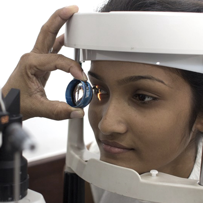
The refraction test is an eye exam that measures how the light is bent and focused on the nerve layer (retina) in the eye. If the image is properly focused on the retina the refraction test becomes zero or Plano and the vision should read as 6/6 ( the line a normal person should read at a distance of 6 meters. )

If the image is not focused properly on the retina then we call it as a refractive error. Then the vision becomes less than 6/6.

The refraction test is done to focus this image back on the retina and gives the power of lenses needed to correct it as a prescription for eyeglasses or contact lenses.

There are four basic refractive errors namely,

Astigmatism (abnormally curved cornea causing blurred vision)

Hyperopia (farsightedness)

Myopia (nearsightedness)

Presbyopia (inability to focus on near objects that develops with age)


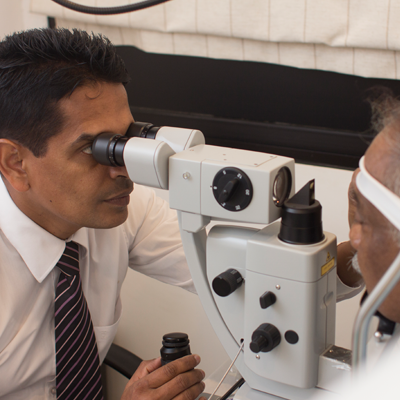
If  the vision can not be improved to the normal values following a refraction test may be due to a complex refractive error ( keratoconus) , some other pathology within the eye ( cataract ) or due to non ocular cause ( stroke).

Please discuss with the doctor for the reason Where your refraction test does not make your vision normal.

K-Reading

Measuring the curvature of the anterior surface of the cornea, particularly for assessing the extent and axis of astigmatism.(abnormally curved cornea causing blurred vision)

Corneal Thickness
  • To find out the healthiness of cornea
  • To get an idea about the accuracy of the eye pressure

Corneal thickness is important because it can mask an accurate reading of eye pressure, causing doctors to treat you for a condition that may not really exist or to treat you unnecessarily when are normal.

  • To plan eye surgeries


Specular Microscopy
  • Specular Microscopy is a noninvasive photographic technique that facilitates rapid and accurate diagnosis of corneal endotheliopathy. (The innermost cell layer of the cornea )
Anterior Segment photography
  • The photographs taken with special equipments to document the diseased conditions of the front part of the eye and to monitor its progression.
OCT - Optical Coherence Tomography

Optical Coherence Tomography is a noninvasive imaging technology used to obtain high resolution cross-sectional images of the retina. The layers within the retina can be differentiated and retinal thickness can be measured to aid in the early detection and diagnosis of retinal diseases and conditions.

OCT testing has become a standard of care for the assessment and treatment of most retinal conditions.

OCT uses rays of light to measure retinal thickness. No radiation or X-rays are used in this test, an OCT scan does not hurt and it is not uncomfortable.

Fundus Retinal Photography

Fundus Retinal Photography uses a fundus camera to record color images of the condition of the interior surface of the eye, in order to document the presence of disorders and monitor their change over time.

The retina is imaged to document conditions such as diabetic retinopathy, age related macular degeneration, macular edema and retinal detachment.

FFA - Fundus Fluorescein Angiography

FFA – Fundus Fluorescein Angiography is a common procedure that is performed to give your doctor more information about the condition of the back of your eye. A small amount of yellow fluorescein dye will be injected into a vein in your arm. The dye travels to your eye where it highlights the blood vessels. It is particularly useful in showing leaking blood vessels and highlighting where the blood supply at the back of the eye is poor. Photographs will then be taken of the eye.It is particularly useful in the management of diabetic retinopathy and macular degeneration.

  • null

    State of the art machinery

Laser

  • null

    Modern Laser Equipment

YAG
Peripheral
PI
SLT
SLT Laser
FLT
PRP
Suture Lysis

There are 4 main laser qualities that is used in medicine. They are

  1. Photo Coagulation
  2. Photo Disruption
  3. Photo Ablation
  4. Photo Evaporation

Out of those 4 qualities,  the first 2 are commonly used in related to eye conditions.

Photo Coagulation

is where laser is used to increase the temperature of a tissue to around 60 degrees and will make the proteins within the cells to coagulate. Eg: Grid laser, Peripheral laser, PRP,

Photo Disruption

Laser energy is used to de stabilize the electrons in the molecules. As a result it becomes unstable which will make the tissues to break up

Eg: YAG, PI, SLT, FLT, Suture Lysis

Electrophysiological Studies

EOG

This test measures the electrical changes within one layer of the retina which occur during dark-adaptation and light-adaptation. The EOG is used to detect and differentiate among several types of retinal dystrophies.

ERG (Flash ERG) & (Pattern ERG)

This test measures the electrical changes within one layer of the retina which occur during dark-adaptation and light-adaptation. The EOG is used to detect and differentiate among several types of retinal dystrophies.

VEP

A visual evoked potential (VEP) is an electrophysiological test that is designed to measure the function of the optic nerves up to the visual cortex.  Patients are seated in a comfortable chair while 5 scalp electrodes are placed on the patient’s head. Then the eyes are stimulated with a light. The resulting electric impulse is grabbed by the those electrodes and printed on a graph which is interpreted by the consultant electrophysiologist.

No pain is involved in this process.

Electrophysiologic testing is useful in diagnosing a variety of inherited retinal and optic nerve diseases, toxic drug exposure, inflammatory conditions, and retinal vascular occlusions.

The primary objective of the electrophysiologic examination is to assess the function of the visual pathway from the photoreceptors of the retina to the visual cortex of the brain. Information obtained from these diagnostic tests helps establish the correct diagnosis or may rule out related ophthalmic diseases.

Electrophysiologic testing is useful in diagnosing a variety of inherited retinal and optic nerve diseases, toxic drug exposure, inflammatory conditions, and retinal vascular occlusions.

Surgical Procedures

Cataract

A Cataract is when the vision is damaged as the result of the natural lens in the human eye losing its transparent property.


Symptoms of Cataract

Double or blurred vision

Sensitivity to light and glare

Less vivid perception of colour


Cataract surgery is performed with a latest technique called phacoemulsification. (Use of a machine to extract the cataract from a small incision)

Advantages of Phacoemulsification

Surgery is done under topical anesthesia (no injections)

Smaller incision (a cut of 1.8 – 2.6 mm)

Use of foldable lens based on latest technology

No sutures

Faster recovery

Corneal Grafts

Corneal transplantation, also known as corneal grafting, is a surgical procedure where a damaged or diseased cornea is replaced by donated corneal tissue.

TPPV (Vitrectomy) Surgery

Vitrectomy is the surgical removal of the vitreous gel from the middle of the eye. It may be done under following conditions.

when there is a retinal detachment

When there is blood in the vitreous gel (vitreous hemorrhage)

During a vitrectomy, the surgeon inserts small instruments into the eye, cuts the vitreous gel, and suctions it out. After removing the vitreous gel, the surgeon may treat the retina with a laser (photocoagulation), cut or remove fibrous or scar tissue from the retina, flatten areas where the retina has become detached, or repair tears or holes in the retina or macula.

At the end of the surgery, silicone oil or a gas bubble is injected into the eye to lightly press the retina against the wall of the eye.

Oil cannot be absorbed by the body, so if an oil bubble is used, you’ll need a second procedure to remove the oil after the retinal detachment has healed.

Collagen Cross-Linking

Corneal collagen cross-linking is a treatment strategy for progressive Keratoconus.This is used in an attempt to make the cornea stronger with riboflavin (vitamin B2) and UV-A light (also known as CXL, C3-R, CCL and KXL), better known as crosslinking. As a first-line treatment the greatest aim of cross-linking is to reduce and stop Keratoconus in the early phase of the condition, and to treat the progressive vision loss that occurs which can lead to corneal transplantation.

Oculoplastic surgery

Oculoplastic surgery includes a wide variety of surgical procedures deals with the Eye socket, eyelids, tear ducts and the face and reconstruction of the eye and associated structures. . They are

  • Ocular reconstructive surgery
  • aesthetic eyelid surgery
  • facial plastic surgery
  • cosmetic surgery
Glaucoma Surgery (Trabeculectomy)

Trabeculectomy is a surgical operation which lowers the intraocular pressure inside the eye (IOP) in patients with glaucoma. This is achieved by making a small hole in the eyewall (sclera), covered by a thin trap-door in the sclera. The fluid inside the eye (known as aqueous humour,) drains through the trap-door to a small reservoir just under the eye surface, hidden by the eyelid. The trap-door is sutured in a way that prevents the fluid inside the eye (aqueous humour) from draining too quickly. By draining aqueous humour the trabeculectomy operation reduces the pressure on the optic nerve and prevents or slows further damage and further loss of vision in glaucoma.

Control of the eye pressure with a trabeculectomy will not restore vision already lost from glaucoma.

Glaucoma is often caused by high pressure inside the eye. Trabeculectomy reduces the eye pressure by draining aqueous humour from the eye.

Retinal Detachment Surgery

Retinal detachment repair is eye surgery to place a retina back into its normal position. The retina is the light-sensitive tissue in the back of the eye. Detachment means that it has pulled away from the layers of tissue around it. Most retinal detachment repair operations are urgent. A detached retina does not get a supply of oxygen. This causes the cells in the area to die, which can lead to blindness.

Squint Correction

Squint surgery is surgery on the extraocular muscles to correct the misalignment of the eyes.

Corneal Surgery

A cornea transplant is an operation used to remove all or part of a damaged cornea and replace it with healthy corneal tissue from the eye of a suitable donor. A cornea transplant is often referred to as keratoplasty or a corneal graft. It can be used to improve sight, relieve pain and treat severe infection or damage.